


The Best Nose Job Transformation
30 June 2025
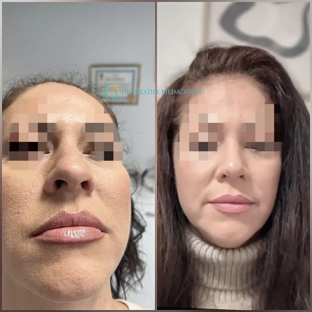
Hispanic Ethnic Rhinoplasty in Turkey
17 October 2025The inverted-V deformity is one of the most common aesthetic complications after rhinoplasty. It appears as a visible indentation on the nasal dorsum following dorsal hump removal, creating a characteristic “inverted V” shape. Not only does this deformity affect the aesthetic outcome, but it can also compromise nasal valve function. In this article, we’ll explore the underlying causes of inverted-V deformity, its anatomical mechanisms, and how to effectively prevent it during primary rhinoplasty.

What Is Inverted-V Deformity?
Inverted-V deformity occurs when the upper lateral cartilages (ULCs) lose their support. This typically happens after the removal of the dorsal hump, causing the cartilages to collapse inward. It presents as a step-off or depression between the nasal bones and cartilaginous dorsum, resulting in a noticeable contour disruption. The cephalic edges of the ULCs become visible under the skin, especially in patients with thin skin, and give the appearance of a sharp angle at the keystone area.
Why Does It Happen? The Anatomic Insight
Inverted‑V deformity appears after rhinoplasty when the upper lateral cartilages (ULCs) lose support and fall inward, revealing a step-like dip on the nasal bridge. Normally, ULCs connect to the nasal bones through a dynamic “T‑frame” structure. When the cartilaginous hump is removed, this T‑frame is broken, leaving the ULCs unsupported. Without that support, the ULCs shift backward, upward, and inward-creating the inverted‑V appearance . This deformity isn’t just aesthetic. As the ULCs collapse, the internal nasal valve narrows, which can interfere with breathing.
In simple terms:
The bony and cartilaginous rooftop becomes disjointed.
The upper lateral cartilages lose tension, drifting inward and creating the V‑shaped indentation
This deformity commonly appears in revision rhinoplasty cases, where previous surgeries may have removed too much cartilage or weakened structural supports
How to Tell If You Have an Inverted‑V Deformity
Ever notice a visible dip or angular line on your nasal bridge after a nose job? That’s often an inverted‑V deformity – a telltale sign your middle vault (the area where nasal bones meet upper lateral cartilages) has collapsed. From the front view, it looks like the letter V, but flipped upside down: two descending lines from the top center of your nose create sharp shadows or depressions on each side.

You may also see:
– Pinched or narrowed bridge, especially in thin‑skinned noses
– Visible bony edges where the cartilage roof
– Breathing issues, due to the collapse narrowing the internal nasal valve
If you think you have an inverted‑V deformity, placing your finger along the bridge may reveal a groove -this often confirms the collapse.
Consulting with a rhinoplasty specialist is the best way to learn about deformities.
Technical Nuances to Improve Outcomes For Experts
It’s important to apply caudal traction early in the reconstruction, before placing grafts or flaps. Don’t suture the ULCs at the very caudal end – this could interfere with their natural divergence near the scroll area. Use a 5.0 PDS suture to apply the correct amount of traction. It can be removed later if tension is maintained structurally or left in place until scar formation. Ensure the roof of the nasal dorsum is restored, whether with cartilage grafts, flaps, diced cartilage in fascia, or T-piecereconstruction.
Management of Nasal Valve Integrity and Structural Support in Primary and Revision Rhinoplasty
If the caudal end of the upper lateral cartilage (ULC) is damaged during lateral osteotomies, the nasal bones may fracture inward (infracture), causing the nasal valve area to shift toward the septum and be affected. This not only narrows the airway size but also disrupts the nasal valve area, potentially leading to collapse in the valve region during normal breathing. During lateral osteotomy at the level of the piriform aperture, near the attachment point of the inferior turbinate, it is beneficial to preserve a caudal piece of nasal bone. When this caudal nasal bone segment is preserved, the patency of the airway and the integrity of the valve area remain intact.
Removal of the bony hump on the nasal dorsum generally has little effect on nasal airway resistance, since this procedure involves the upper part of the nasal airway and does not interfere with airflow. However, when reducing the cartilaginous portion of a dorsal hump, caution is necessary because excessive manipulation or resection of the ULCs can adversely affect the nasal valve area, leading to collapse and increased nasal resistance. Scar contracture or adhesions in the nasal valve area can cause severe airway obstructions, resulting in nasal blockage that is difficult to correct. When the ULCs curve inward toward the septum, stability can be restored by using spreader grafts, medial upper lateral “turn-in” grafts, or by suturing the ULC back to the septum.
Resection or weakening of the alar cartilages (ALC) can lead to alar collapse during inspiration. If the ALCs must be incised and separated, the resected ends should be sutured together, and sometimes an alar onlay graft may be required for additional support. The continuity of the lateral crural strip should be preserved. Reduction surgery of the lower one-third of the nose can damage the interdomal ligament (intercartilaginous membrane) and compromise the integrity of the nasal valve area.
In cases of a pinched middle third of the nose or nasal valve collapse with airway obstruction, bilateral spreader grafts can be used. The cartilage grafts are positioned between the dorsal margin of the ULC and the nasal septum. This maneuver widens the middle third of the nose and opens the airway by separating the upper lateral cartilage from the septum.

Bilateral spreader grafts. A. Pinched upper lateral cartilages (ULCs) giving a narrow appearance to the middle third of the nose. B. Placement of spreader grafts between the cartilage graft and the nasal septum. C. Placement of bilateral spreader grafts and correction with a widened appearance of the middle third
The Role of Muscle Tension and Fascia
Beyond cartilage and bone, nasal muscle tone and surrounding fascia also contribute to dorsum stability. Maintaining the integrity of the scroll area and associated ligaments can support ULC positioning, especially in dynamic facial movement.
Surgeons are advised to preserve these structures whenever possible, or to reattach them if dissected during the procedure.
How Caudal Traction Prevents the Deformity
The most effective preventive technique observed was caudal traction-pulling the ULCs downward (toward the septum) during reconstruction. This maneuver restores their tension and brings them back into contact with the nasal bones, recreating a smooth transition and preventing the visible step-off.
For long-term success, the ULCs must be fixed in this position using spreader grafts, spreader flaps, or by reconstructing the T-frame. Scar formation over time will help stabilize the new configuration.
Important note: Caudal traction is only effective in primary rhinoplasty. In revision cases, scar tissue prevents the ULCs from responding to this maneuver.

What to Expect from Revision Rhinoplasty to Correct Inverted V- Deformity
- Full structural assessment
- Your surgeon will visually and physically evaluate the nose—palpating cartilage, inspecting skin thickness, and checking valve function. Imaging may also be used to assess bone alignment and internal airway .
- Open or closed surgical approach
- Though some surgeons use a closed technique, most prefer the open approach for revisions—it offers better visibility and precision during complex reconstructions.
- Cartilage harvesting
- TIf the available septal cartilage is insufficient, the surgeon will probably require additional grafts taken from the ear or rib.
- Recovery Expectations
- Duration: Revision procedures typically last 4 hours, followed by 5–8 days of initial recovery with splints and cast.
- Swelling/Bruising: Expect noticeable but temporary swelling for 2–3 weeks; most activity is possible after 2 weeks.
- Final results: To see the final result, you will have to wait at least one year, since this period is needed for the cartilage grafts to fuse, for the swelling to go away, and for the new structure to take root and settle.
- Breathing improvements: Aside from cosmetic effects, many patients report significantly improved airflow due to restored valve support .
Revision rhinoplasty carries complications and risks, including infection, bleeding, scarring, or graft problems, especially if you have had previous surgeries. Complication rates lie between 8–15%, though most are minor.
Final Conclusions
Surgical success relies heavily on surgeon experience, solid pre-op planning, and clear patient communication about expectations. The best correction of the ‘inverted V deformity’ is surgical revision rhinoplasty with structural cartilage grafts and possible bone reshaping. Proper evaluation and treatment from an experienced rhinoplasty specialist, as Dr. Abulkadir Kilimcioglu, can restore not just the shape but also the breathing capacity of your nose.
If you’re planning a revision, seek a surgeon who prioritizes not just aesthetics but also internal nasal anatomy.
